


Evaluation and Treatment Approaches: Giant Cell Tendon Sheath Tumor
1. Introduction
The purpose of this study is to examine the evaluation and treatment approaches for giant cell tendon sheath tumors. The pathophysiology, clinical features, diagnostic methods, imaging findings, treatment options and rehabilitation processes of the tumors will be discussed.
2. Giant Cell Tendon Sheath Tumor: Definition and Epidemiology
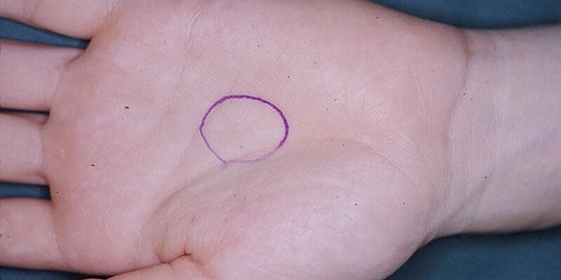
Giant cell tendon sheath tumor is one of the most common benign tumors of the musculoskeletal system. Originating from tendinous tissue, this tumor usually occurs in young adults and during adolescence. It is slightly more common in women than in men. It is usually found in extremity limbs such as the wrist and ankle.
Epidemiological studies show that the frequency of this tumor has increased over the years. In addition, an increase in the number of cases of giant cell tendon sheath tumors detected by radiological examinations is observed.
3. Pathophysiology and Etiology
The pathophysiology of giant cell tendon sheath tumors is generally not fully understood, but it is known that the tumor is benign in most cases. Etiological factors are as follows:
- Genetic predisposition
- Chronic irritation of tendon tissue
- Traumatic injuries
- Hormonal factors
- Inflammatory processes
Further research is needed on the pathophysiology and etiology of the tumor.
4. Clinical Findings and Diagnostic Methods
Giant cell tendon sheath tumor usually presents with the following symptoms:
- Pain
- Swelling
- Localized tenderness
Diagnosis is usually made with the following methods in addition to clinical findings:
- Radiography: To determine tumor borders and calcification.
- Magnetic Resonance Imaging (MRI): To assess tumor size and extent of spread.
- Computed Tomography (CT): For detailed imaging of bone and soft tissues.
- Biopsy: Used as the gold standard for definitive diagnosis.
5. Imaging and Laboratory Findings
Imaging methods play an important role in the diagnosis of giant cell tendon sheath tumors:
- Radiography: To determine the tumor borders and calcification.
- Magnetic Resonance Imaging (MRI): Provides detailed information about the tumor size, degree of spread and its relationship with surrounding tissues.
- Computed Tomography (CT): Used for detailed imaging of bone and soft tissues.
Biopsy provides definitive diagnosis as a result of histopathological examination.
6. Treatment Options and Approaches
Treatment methods for giant cell tendon sheath tumors vary depending on the location and size of the tumor. Treatment options include:
- Surgical Intervention: It is performed to completely remove the tumor.
- Monitoring and Follow-up: Regular follow-up is required to minimize the risk of recurrence after surgery.
7. Rehabilitation and Prognosis
Post-treatment rehabilitation plays an important role in the recovery process of patients. Physiotherapy programs include the following goals:
- Restoring joint range of motion
- Increasing muscle strength
- Returning to daily life activities
Prognosis: Early diagnosis and treatment can usually yield good results. However, the risk of recurrence should be considered and patients should be followed up regularly.
After completing his high school education, Dr. Özgür Erdoğan successfully completed Istanbul University Faculty of Medicine and received his medical doctor title. He completed his higher specialization at Haydarpaşa Numune Training and Research Hospital Orthopedics and Traumatology Clinic. In March 2021, he received the title of Associate Professor of Orthopedics.


